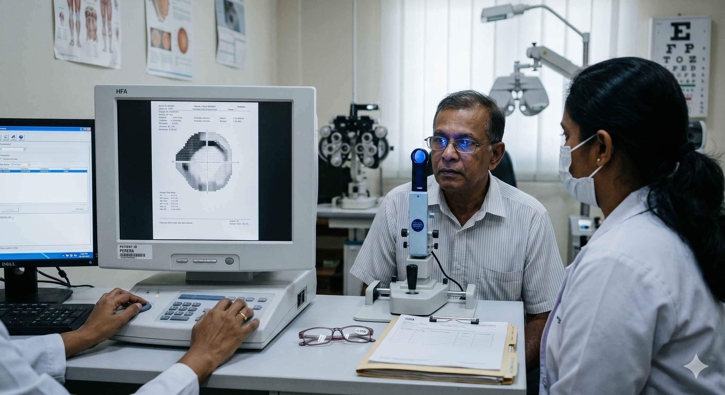
Case 1
Mr. Perera walks into the clinic for a routine check-up. He hasn’t seen an eye doctor in over five years but recently decided to visit because he felt his “reading glasses weren’t working as well as they used to.“
History:
- Chief Complaint: Gradual, painless blurring of vision, primarily noticed when reading or driving at night.
- History of Presenting Illness: He denies any sudden loss of vision, eye pain, or seeing halos around lights. When questioned closely about his peripheral vision, he mentions, “I feel like I’ve become a bit more clumsy lately, bumping into the doorway once or twice, but I thought it was just my age.”
- Past Medical History: Managed for hypertension for 10 years; no history of diabetes.
- Past Ocular History: No history of eye trauma or surgery. He uses over-the-counter reading glasses (+2.25D).
- Family History: Significant. His elder brother was diagnosed with “high eye pressure” and uses daily eye drops.
- Social History: Non-smoker, enjoys gardening and reading.
Clinical Examination
Upon examination, the following findings are noted:
- Visual Acuity (VA)
- Right Eye (OD): 6/9 (Snellen), improving to 6/6 with pinhole.
- Left Eye (OS): 6/9 (Snellen), improving to 6/6 with pinhole.
- Near Vision: N8 with his current correction.
- Intraocular Pressure (IOP) – Goldmann Applanation Tonometry
- OD: 24 mmHg
- OS: 26 mmHg
(Normal range is typically 10–21 mmHg).
-
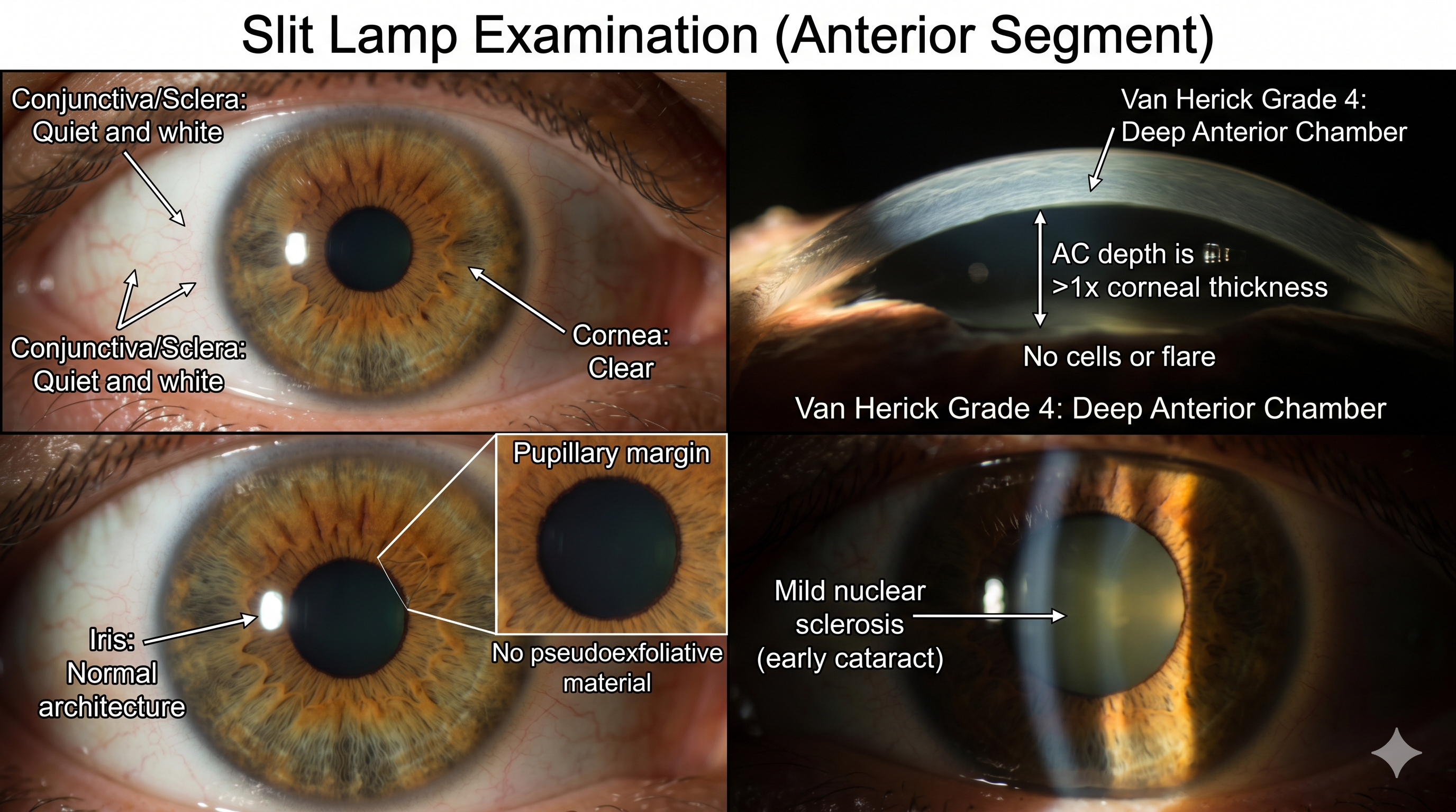
Slit Lamp Examination (Anterior Segment)
- Conjunctiva/Sclera: Quiet and white.
- Cornea: Clear, no signs of pigment on the endothelium (Krukenberg spindle).
- Anterior Chamber: Deep and quiet (Van Herick Grade 4). No cells or flare.
- Iris: Normal architecture, no pseudoexfoliative material on the pupillary margin.
- Lens: Mild nuclear sclerosis (early cataract) bilaterally.
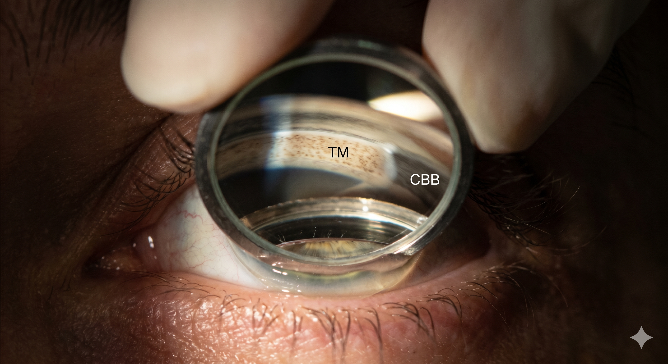
- Gonioscopy
- Both Eyes: Angles are open to the ciliary body band (Grade 4 Shaffer) in all four quadrants. There is normal trabecular pigmentation and no peripheral anterior synechiae (PAS).
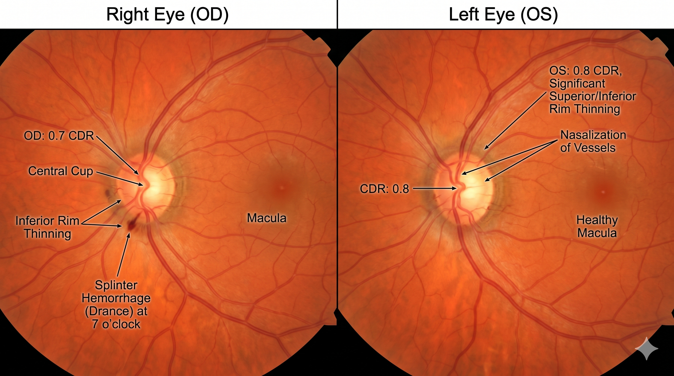
- Posterior Segment (Fundoscopy)
The most critical findings are at the optic nerve head:
- Optic Disc OD: Large cup-to-disc ratio (CDR) of 0.7. Noted thinning of the inferior neuroretinal rim and a small splinter hemorrhage (Drance hemorrhage) at the 7 o’clock position.
- Optic Disc OS: CDR of 0.8. Significant thinning of the superior and inferior rims (vertical elongation of the cup). Noted “nasalization” of the vessels.
- Macula/Periphery: Healthy in both eyes; no signs of hypertensive retinopathy.